Posts

Showing posts from May, 2024

VASOSPASM AFTER SUBARACHNOID HEMORRHAGE
Image
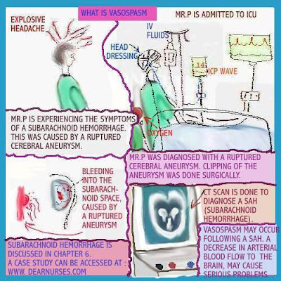
  Subarachnoid Hemorrhage   The image above shows a patient displaying the symptoms of Subarachnoid  hemorrhage. A CAT scan  was done to confirm the diagnosis.   Surgical intervention was done, for clipping of the aneurysm. He will be cared for in the ICU. VASOSPASM  This condition may occur following a Subarachnoid hemorrhage. TCD ( Transcranial Doppler) ultrasound is ordered by the doctor, following a Subarachnoid hemorrhage. This is a noninvasive study to evaluate    blood flow in the brain.
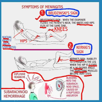
BRUDZINSKI'S AND KERNIG'S SIGNS

Image
  Brudzinski's and Kernig's signs  are seen in patients who have Meningitis or  Subarachnoid hemorrhage. Brudzinski's sign  -When the examiner flexes the patient's neck , the knees and  hips flex at the same time. Kernig's sign  - inability to straighten the leg when the hip is flexed at a 90 degree angle. The hamstring muscles become very stiff.

FETAL HEART MONITORING

Image
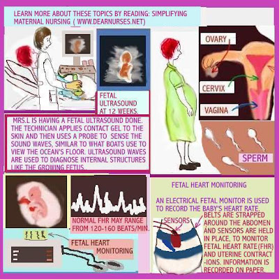
                                                                Fetal Heart Monitoring  Fetal Ultrasound In the image above, a fetal ultrasound is being done, by a technician. Contact gel is applied to the skin , followed by the use of a probe, to sense sound waves. This technique is similar to what boats use, to view the ocean's floor. Ultrasound waves are used to diagnose internal structures like the growing fetus. Learn more about Placenta previa Addressing fetal heart monitoring Fetal heart monitoring is done to monitor the baby's heart rate. An electric fetal monitor is used for this purpose. Belts are strapped around the abdomen and sensors are held in place, to monitor FHR ( fetal heart rate)and uterine contractions.   Information is recorded on paper.

ADDRESSING PRETERM LABOR

Image
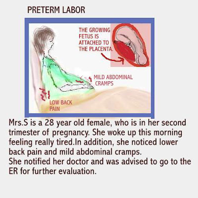
Scenario:  Mrs. S is a 28 year old who is in her second trimester of pregnancy. She woke up feeling very tired. In addition, she noticed lower back pain and mild abdominal cramps. She notified her doctor and was advised to go to the  ER for further evaluation. Preterm labor is a very common occurrence in pregnancy. lts causes are not clearly understood. It may be triggered by events such as stress, trauma,  heavy lifting or infection.  Signs and symptoms include:  Premature contractions,  abdominal cramping, low back  pain and vaginal bleeding . Patients should be encouraged  to go to their doctor or  Emergency Room  if necessary.  Close monitoring of the patient in preterm labor is done. Medications such as Calcium channel blockers, Tocolytics ( Terbutaline and Albuterol) , antibiotic therapy to treat the infection,  Magnesium Sulfate and Prostaglandins are used in treating this condition.

THIRD DEGREE HEART BLOCK AND LETHAL RHYTHMS

Image
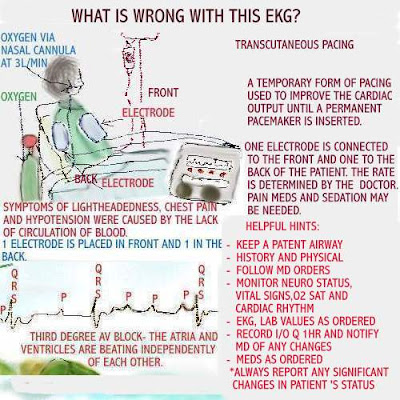
                                                       Third degree AV block The patient in third degree heart block is likely to display  the EKG rhythm seen above. Typically, in a healthy heart , the atria contract at the same time, forcing blood into the ventricles. This is followed by the ventricles contracting,  at the same time, pushing blood out of the heart. In third degree heart block, because the heart is damaged, it cannot pump as effectively as a healthy heart. This causes the atria and ventricles to beat independently of each other.  The P wave may not be followed by the QRS. The EKG tracing above demonstrates this. This means blood flow is lacking. Symptoms  include: chest pain, feeling lightheaded and  hypotension. Causes of third degree heart block: - cardiomyopathy - coronary artery disease - surgical intervention - medications such as beta blockers,  calcium channel blockers and Digoxin Treatment may vary. AHA has guidelines in place for the course treatment. Oxygen