THIRD DEGREE HEART BLOCK AND LETHAL RHYTHMS
Third degree AV block
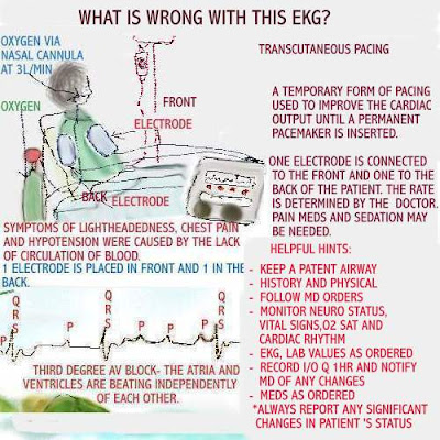
Third degree AV block
The patient in third degree heart block is likely to display
the EKG rhythm seen above. Typically, in a healthy heart,
the atria contract at the same time, forcing blood into the
ventricles. This is followed by the ventricles contracting,
at the same time, pushing blood out of the heart.
the EKG rhythm seen above. Typically, in a healthy heart,
the atria contract at the same time, forcing blood into the
ventricles. This is followed by the ventricles contracting,
at the same time, pushing blood out of the heart.
In third degree heart block, because the heart is damaged,
it cannot pump as effectively as a healthy heart. This causes
the atria and ventricles to beat independently of each other.
it cannot pump as effectively as a healthy heart. This causes
the atria and ventricles to beat independently of each other.
The P wave may not be followed by the QRS. The EKG
tracing above demonstrates this. This means blood flow
is lacking.
tracing above demonstrates this. This means blood flow
is lacking.
Symptoms include: chest pain, feeling lightheaded and
hypotension.
hypotension.
Causes of third degree heart block:
- cardiomyopathy
- surgical intervention
- medications such as beta blockers,
calcium channel blockers and Digoxin
Treatment may vary. AHA has guidelines in place for the
course treatment. Oxygen will be required. Atropine may be
used first depending, on how stable the patient is. The doctor
may choose transcutaneous pacing first and later, have a
course treatment. Oxygen will be required. Atropine may be
used first depending, on how stable the patient is. The doctor
may choose transcutaneous pacing first and later, have a
permanent pacemaker put in.
Documentation of all interventions, is important. Please
read the image above, for more helpful information.
read the image above, for more helpful information.
Lethal rhythms

***American Heart Association has protocols in place for each
EKG rhythm that requires immediate intervention.
EKG rhythm that requires immediate intervention.
There are some EKG rhythms that are considered to be lethal.
Instant intervention is necessary for good outcomes.
Here are the most lethal rhythms:
Asystole - sometimes referred to as " flat-line. The electrical
heart's conduction system is not functioning. There are no
ventricular contractions. The patient is pulseless and
unresponsive.
ventricular contractions. The patient is pulseless and
unresponsive.
PEA - the patient in PEA will have a heart rhythm, but is
unresponsive. This may be caused by hypovolemia, cardiac
tamponade, pneumothorax, drug overdose, thrombosis,
unresponsive. This may be caused by hypovolemia, cardiac
tamponade, pneumothorax, drug overdose, thrombosis,
and trauma.
Ventricular Tachycardia - There may or may not be a pulse
present. The ventricles are beating rapidly and there is no
effective blood flow. Ventricular Tachycardia without a pulse
is treated the same as ventricular fibrillation. Typically the
patient will be unresponsive and require immediate CPR
and defibrillation.
present. The ventricles are beating rapidly and there is no
effective blood flow. Ventricular Tachycardia without a pulse
is treated the same as ventricular fibrillation. Typically the
patient will be unresponsive and require immediate CPR
and defibrillation.
Ventricular Fibrillation - There is a chaotic rhythm and no
atrial activity. The ventricles are quivering , so there is no
cardiac output. There is no pulse and the patient may become
unresponsive. Immediate action to defibrillate is usually done.
atrial activity. The ventricles are quivering , so there is no
cardiac output. There is no pulse and the patient may become
unresponsive. Immediate action to defibrillate is usually done.


Comments
Post a Comment